Viral culture tools form the foundation of modern virology research and diagnostic work. These specialized instruments and materials allow scientists to grow, isolate, and study viruses in controlled laboratory settings. Without reliable viral culture tools, researchers would struggle to develop vaccines, identify new pathogens, or understand how viruses replicate and spread.
This guide covers the essential equipment, techniques, and applications that define viral culturing today. Whether a lab is studying influenza strains or working on emerging infectious diseases, the right viral culture tools make the difference between success and failure.
Table of Contents
ToggleKey Takeaways
- Viral culture tools are essential equipment and materials used to grow, isolate, and study viruses in controlled laboratory settings.
- Core viral culture tools include CO2 incubators, biosafety cabinets, inverted microscopes, centrifuges, and cryogenic storage systems.
- Cell lines like Vero, MDCK, and HeLa serve as living substrates where viruses replicate, with each supporting different virus types.
- Common culturing techniques include monolayer culture, suspension culture, plaque assays, shell vial culture, and embryonated egg culture.
- Viral culture tools support critical applications such as vaccine development, antiviral drug screening, clinical diagnostics, and basic virology research.
- The quality and maintenance of viral culture tools directly impact experimental outcomes and research success.
What Are Viral Culture Tools?
Viral culture tools are the equipment, supplies, and biological materials used to propagate viruses outside their natural hosts. Unlike bacteria, viruses cannot reproduce on their own. They require living host cells to replicate. This fundamental characteristic shapes every aspect of viral culturing.
The basic components of viral culture tools include:
- Host cell systems (cell lines, primary cells, or embryonated eggs)
- Growth media and supplements to maintain cell viability
- Incubators with precise temperature and CO2 control
- Biosafety cabinets for sterile handling
- Microscopes for monitoring cell health and viral effects
Viral culture tools also encompass consumables like flasks, plates, pipettes, and cryogenic storage equipment. Each piece plays a specific role in keeping cells alive while viruses replicate within them.
The quality of viral culture tools directly impacts experimental outcomes. Contaminated media or failing incubators can ruin weeks of work. Labs invest significant resources in maintaining and calibrating their viral culture tools to ensure consistent results.
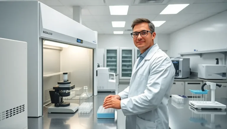
Key Equipment for Viral Culturing
Successful viral culturing depends on several core pieces of equipment working together. Each serves a distinct purpose in the workflow.
CO2 Incubators maintain the temperature (typically 37°C) and carbon dioxide levels (usually 5%) that mammalian cells need to survive. Modern incubators feature HEPA filtration and copper-lined interiors to reduce contamination risk.
Biosafety Cabinets protect both researchers and samples. Class II cabinets are standard for most viral work, providing HEPA-filtered air flow that keeps pathogens contained while maintaining a sterile work surface.
Inverted Microscopes let scientists observe cells growing in flasks and plates. Phase contrast optics reveal cell morphology without staining, making it easy to spot the cytopathic effects viruses cause.
Centrifuges separate viral particles from cell debris and concentrate samples for analysis. Refrigerated models preserve viral integrity during processing.
Cryogenic Storage Systems preserve cell lines and viral stocks at ultra-low temperatures (-80°C freezers or liquid nitrogen tanks). Proper storage of viral culture tools and materials ensures labs can maintain their resources for years.
Cell Lines and Growth Media
Cell lines serve as the living substrate where viruses replicate. Common options include Vero cells (derived from African green monkey kidney), MDCK cells (for influenza), and HeLa cells (human cervical cancer cells). Each cell line supports different virus types.
Growth media supply nutrients, amino acids, vitamins, and buffers that cells need. DMEM (Dulbecco’s Modified Eagle Medium) and RPMI 1640 are popular choices. Labs supplement these with fetal bovine serum (FBS) to provide growth factors, though serum-free alternatives are gaining ground.
The interaction between cell lines, media, and viral culture tools determines how well a particular virus will grow. Some viruses are fastidious, they only replicate in specific cell types under narrow conditions. Others grow readily in multiple systems.
Common Viral Culture Techniques
Several established methods dominate viral culturing. The choice depends on the virus type, research goals, and available viral culture tools.
Monolayer Culture involves growing a single layer of cells on flask or plate surfaces. Researchers inoculate the monolayer with virus-containing samples and watch for cytopathic effects, visible changes like cell rounding, detachment, or syncytia formation. This technique works well for many respiratory and enteric viruses.
Suspension Culture keeps cells floating in liquid media rather than attached to surfaces. Some viruses, particularly those infecting immune cells, grow better in suspension systems. Spinner flasks or bioreactors maintain constant gentle agitation.
Plaque Assays quantify infectious virus particles. A cell monolayer is infected, then covered with a semi-solid overlay (agar or methylcellulose). As viruses spread locally, they create clear zones called plaques. Counting plaques gives a measure of viral titer.
Shell Vial Culture accelerates viral detection for clinical diagnostics. Cells grow on coverslips in small vials. After inoculation, centrifugation forces viral attachment, and immunofluorescent staining identifies specific viruses within 24-48 hours.
Embryonated Egg Culture remains important for influenza vaccine production. Virus is injected into the allantoic cavity of fertilized chicken eggs, where it replicates to high titers. This traditional method predates cell culture but still produces most flu vaccines worldwide.
Each technique requires specific viral culture tools and expertise. Labs often maintain capabilities for multiple methods to handle different project requirements.
Applications in Research and Diagnostics
Viral culture tools support a wide range of scientific and clinical activities. Their applications span basic research, drug development, and patient care.
Vaccine Development relies heavily on viral culturing. Scientists must grow large quantities of virus to create inactivated or attenuated vaccines. The COVID-19 pandemic demonstrated how quickly viral culture tools can scale up when global health demands it.
Antiviral Drug Screening uses cultured viruses to test candidate compounds. Researchers infect cells, apply potential drugs, and measure how well they inhibit viral replication. This process identifies promising leads before expensive animal studies.
Clinical Diagnostics sometimes require viral culture to confirm infections or characterize outbreak strains. While molecular methods like PCR are faster, culture provides live virus for additional testing, including antiviral susceptibility profiles.
Basic Research into viral biology depends on culture systems. How does a virus enter cells? What host factors does it hijack? How does the immune system respond? Answering these questions requires viral culture tools that produce consistent, well-characterized viral stocks.
Environmental Monitoring uses viral culture to detect pathogens in water, food, and air samples. Public health labs culture samples to identify contamination sources during outbreak investigations.
The versatility of viral culture tools makes them indispensable across multiple disciplines. From academic labs studying fundamental biology to pharmaceutical companies developing new treatments, these resources enable critical work.